Radiotherapy patient systems from gKteso
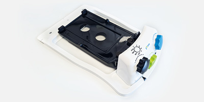
RPS head
The most favourable solution of angle correction in radiotherapy. Better results In combination with several thermal masks.
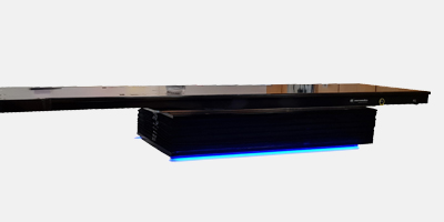
RPS Mini
Multiple brain metastases corrected via XVI Data. The 6 DoF add on couch allows correction after cone beam CT.
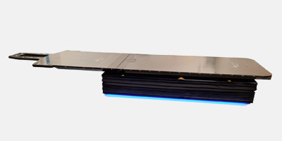
RPS SGRT
Motion management during treatment. Correction of motion during radiation in combination with surface scanner.
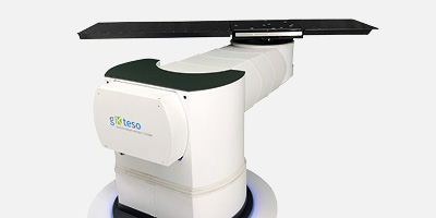
RPS fixedbeam
Innovative solutions in radiotherapy treatment. Open a new direction of cost reduction with gkteso.
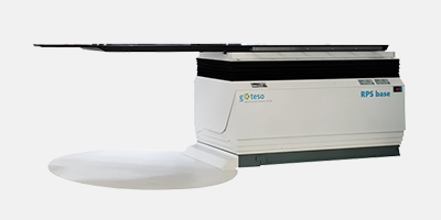
RPS Base
More range for more correction possibilities. Angle correction up to +/- 5 degress on each axis.
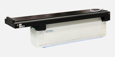
RPS Base MR
– Large longitudinal travel range
– Flexible API
– Kevlar board
gKteso: Solutions for radiotherapy
+49 8234 966 38 41
- 28 years experience
- Safe and economical solutions “outside the box“
- Benfits for Medical Physicists in radiation cancer therapy
Do you have questions about our solutions?
We are looking forward to your inquiry!
Stereotactic radiotherapy
Submillimeter correction
Compatible with various immobilization manufacturer
Stereotactic radiotherapy is usually performed in six steps, which also apply for brain tumor radiotherapy:
• Indication
• Planning: CT for stereotactic radiotherapy
• Radiation plan
• Simulation and verification of the treatment
• Brain tumor radiotherapy
• Follow-up care
Imaging techniques are continuously improving the quality of brain tumor radiotherapy. Even in the planning stage of stereotactic radiotherapy, images from MRI and CT are used to precisely determine the position of the tumor in the patient for radiation. The radiation plan specifies the contouring of the target structure and the doses and frequency of radiation.
A dummy can then be used to simulate and verify the radiotherapy. This is followed by the actual treatment of the immobilized patient in accordance with the established parameters. If the radiotherapy machine images are then evaluated before the radiation treatment and the patient is corrected with the help of a 6D couch, the surrounding tissue is spared as much as possible. The success of the treatment is monitored during follow-up care.
- more precision
- more protection of healthy tissue
Image guided radiotherapy
Improve the stereotactic radiotherapy success rate
Motion under control as best as possible
The advantage of 4D radiotherapy is that the movement of the tumor is incorporated into the treatment, increasing the success rate of stereotactic radiotherapy. The main advantages for patients are that this can reduce the target structure and better protect the surrounding tissue.
- No compromise in treatment
- Every submillimeter count
Brain radiotherapy
Submillimeter correction is so important
Pinpoint radiation for the life beyond
Predefined radiotherapy imaging protocols ensure efficient workflows during planning. This is the only way to ensure direct processing of all the images in the treatment planning system. Brain radiotherapy, in particular, relies on ever new technical improvements to provide patients with the best possible protection against the side effects of radiotherapy. So, in the future for brain radiotherapy, patient biovariability will already be included in the planning stage. A wide variety of imaging techniques are also consulted and considered for planning, because each imaging technique has its specific advantages. To this end, uniform radiotherapy imaging protocols are important for automatically processing the acquired images.
Once all the data have been processed, even lower dose inputs can be achieved in the future during irradiation, which in turn means less stress for patients. This is especially important to consider when sensitive structures, such as the lens of the eye, are in close proximity.
- Compatible for all immobilization devices
- Correct the setup inaccuracies
Fractioned radiotherapy
Use the best thermal masks for radiotherapy for the best immobilization
Our connecting plate enables this feature
Portal imaging in radiotherapy is an important tool for verifying correct patient positioning for fractionated stereotactic radiotherapy. In portal imaging in radiotherapy, images are generated by projecting the inside of the body with the treatment beam onto a screen and these are then made available as a digital image. The procedure supports the correct positioning of the patient, especially for high-precision, fractionated stereotactic radiotherapy. For small-volume tumors especially, fractionated stereotactic radiotherapy is an efficient procedure that allows the surrounding tissue to be spared as much as possible. For this reason, it is also used for brain metastases. A mask for radiotherapy is used to ensure that the patient’s head remains absolutely immobile during high-precision radiotherapy.
The mask for the thermoplastic shell radiotherapy of brain metastases is usually a thermoplastic shell that is customized to fit the patient’s face and head. The thermoplastic shell is heated for this purpose so that the material can be flexibly applied to the contours of the face. After cooling, the thermoplastic tray for the radiotherapy of brain metastases is rigid and reliably secures the head to the treatment couch.
- Flexible connecting plate
- 6D correction
Radiotherapy mask
The right thermal shell is so important for the best treatment
Fit for the future thanks to the compatibility to all immobilization device
Stereotactic radiotherapy in the brain aims to fight the tumor while sparing the patient as much as possible. As a result, patient positioning is critical in radiotherapy: The more precisely the patient is positioned under the linear accelerator, the more accurate the radiotherapy. Immobilization radiotherapy ensures that the body remains in the ideal position for treatment. This is particularly important for stereotactic radiotherapy in the brain, where sensitive structures are located in the immediate vicinity of the tumor. To minimize side effects, patient positioning in radiotherapy is precisely planned and calculated in advance of treatment. Once the ideal position is found, deviations are minimized by immobilization – and radiotherapy can begin.

- Sparing the patient as much as possible.
- The more precisely the patient is positioned under the linear accelerator, the more accurate the radiotherapy.
- To minimize side effects, patient positioning in radiotherapy is precisely planned and calculated in advance of treatment.
